



Microvascular Bone and Soft Tissue Reconstruction
Thank you for your feedback!
How far can reconstructive microsurgery go when bone is infected, fixation has failed, soft tissue is missing, or amputation seems like the only remaining option?
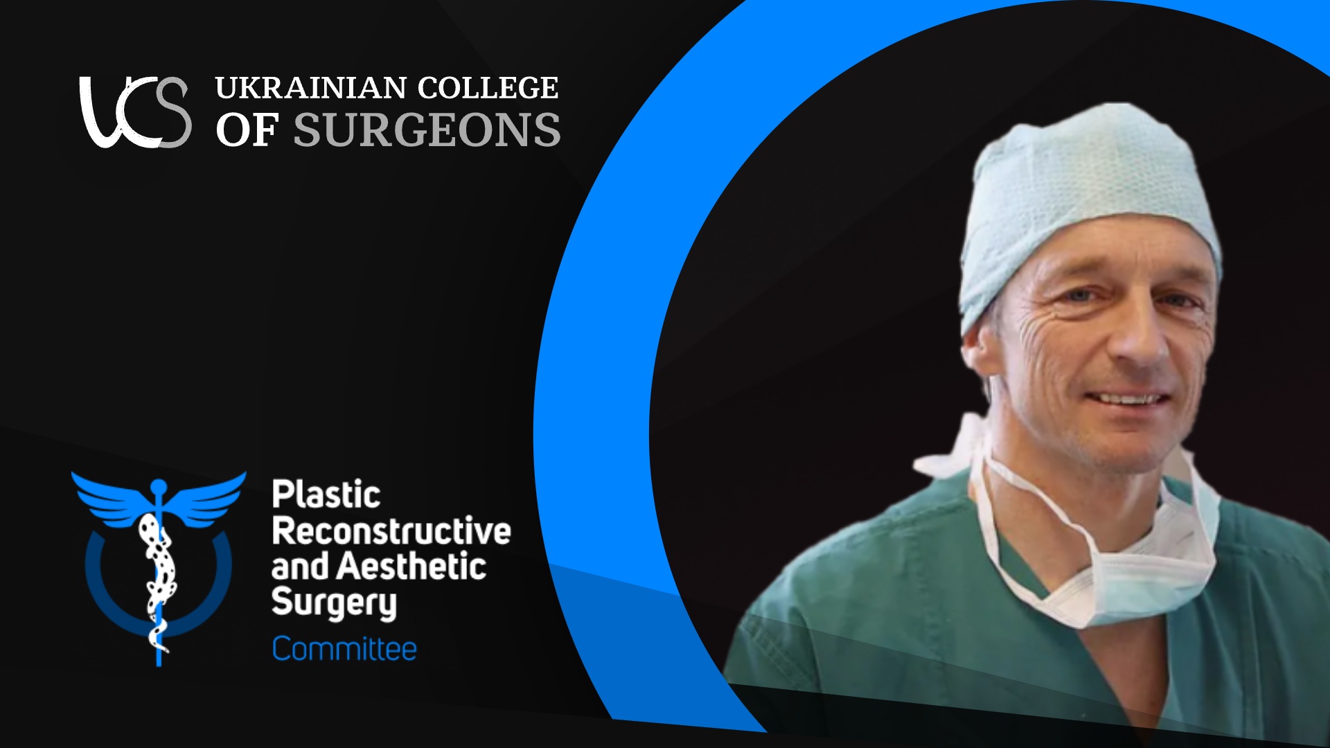
Through a wide range of clinical cases, Dr Heinz Berger explores vascularized bone and soft tissue reconstruction using the medial femoral condyle, iliac crest and fibula flaps, often combined with the ALT flap. The lecture moves from hand and scaphoid reconstruction to massive long-bone defects, heel salvage, tumor-related defects and some highly unusual reconstructive challenges.
A major focus is the biology behind vascularized reconstruction: periosteal perfusion, graft hypertrophy under loading, recipient-vessel selection in scarred or compromised tissue, ICG assessment and anastomoses involving vessels below 0.8 mm. Long-term follow-up cases show what happens years after reconstruction—not only what the immediate postoperative images look like.
The discussion also becomes highly practical: which flaps are useful when starting a microsurgery program, what instruments are actually essential, why combined bone and soft-tissue defects may be better addressed in one stage, and how the approach changes when infection or existing implants are present. The final Q&A connects these principles directly to severe trauma and mine-related calcaneal defects relevant to Ukrainian surgical practice.
Through a wide range of clinical cases, Dr Heinz Berger explores vascularized bone and soft tissue reconstruction using the medial femoral condyle, iliac crest and fibula flaps, often combined with the ALT flap. The lecture moves from hand and scaphoid reconstruction to massive long-bone defects, heel salvage, tumor-related defects and some highly unusual reconstructive challenges.
A major focus is the biology behind vascularized reconstruction: periosteal perfusion, graft hypertrophy under loading, recipient-vessel selection in scarred or compromised tissue, ICG assessment and anastomoses involving vessels below 0.8 mm. Long-term follow-up cases show what happens years after reconstruction—not only what the immediate postoperative images look like.
The discussion also becomes highly practical: which flaps are useful when starting a microsurgery program, what instruments are actually essential, why combined bone and soft-tissue defects may be better addressed in one stage, and how the approach changes when infection or existing implants are present. The final Q&A connects these principles directly to severe trauma and mine-related calcaneal defects relevant to Ukrainian surgical practice.
PRAS-02
Course
Fundamentals of Microsurgical and Free-Flap Reconstruction
 01
1:27:03
Free Flap Surgery: Microsurgical Aspects and Patient ManagementExplore the evolution and application of free flap reconstruction in modern surgery, focusing on complex trauma management and multidisciplinary orthoplastic approaches.
01
1:27:03
Free Flap Surgery: Microsurgical Aspects and Patient ManagementExplore the evolution and application of free flap reconstruction in modern surgery, focusing on complex trauma management and multidisciplinary orthoplastic approaches.
 02
1:20:50 Now playing
Microvascular Bone and Soft Tissue ReconstructionA case-driven lecture on vascularized bone grafts, free flaps and supermicrosurgery for complex trauma, nonunion, infection and limb salvage.
02
1:20:50 Now playing
Microvascular Bone and Soft Tissue ReconstructionA case-driven lecture on vascularized bone grafts, free flaps and supermicrosurgery for complex trauma, nonunion, infection and limb salvage.
 03
48:32
The Path to Microsurgery: Clinical Cases of Reconstruction of Combined Soft Tissue and Bone DefectsA practical account of developing a microsurgical service through complex reconstructive cases, failed flaps, team training, and increasingly demanding limb-salvage procedures.
03
48:32
The Path to Microsurgery: Clinical Cases of Reconstruction of Combined Soft Tissue and Bone DefectsA practical account of developing a microsurgical service through complex reconstructive cases, failed flaps, team training, and increasingly demanding limb-salvage procedures.
 04
1:00:51
Free Flap Planning: From Patient Selection to a Reliable Surgical SystemFree flap surgery is more than microsurgical technique—it is a carefully coordinated system. This lecture explores how planning, team organization, vascular imaging, anesthesia, flap selection and rehabilitation shape the final reconstructive result.
04
1:00:51
Free Flap Planning: From Patient Selection to a Reliable Surgical SystemFree flap surgery is more than microsurgical technique—it is a carefully coordinated system. This lecture explores how planning, team organization, vascular imaging, anesthesia, flap selection and rehabilitation shape the final reconstructive result.
 Microvascular Bone and Soft Tissue Reconstruction